

Getting an ultrasound test is one of the things that most thrills and terrifies you when you are pregnant. One of the most important aspects of prenatal care throughout pregnancy ultrasound. Ultrasound scans are performed to evaluate the fetus’s growth and development, track the mother’s and child’s health, and identify any possible issues. However, many soon-to-be parents may find an ultrasound report to be confusing and overwhelming.
We’ll give you a thorough explanation of how to read pregnancy ultrasound report, in this post, along with information on the many kinds of ultrasound scans, what to expect during the scan, and how to interpret the results. You will know exactly what each area of the prenatal ultrasound report signifies by the end of this article, along with how to discuss the results with your healthcare physician.
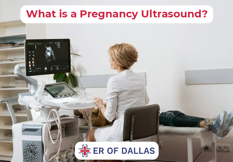
What is a Pregnancy Ultrasound?
During a pregnancy ultrasound, the mother’s reproductive organs and the growing fetus are imaged using high-frequency sound waves. With every pregnancy, the typical number of ultrasounds varies.
Sonograms, commonly known as ultrasounds, are used to screen for any issues and keep an eye on a fetus’s healthy development. A fetal echocardiogram, or detailed ultrasound of the fetus’s heart, is one of the more sophisticated ultrasounds available in addition to the normal ultrasound. Other advanced ultrasounds include 3-D and 4-D ultrasounds.
A transducer is a device that sends sound waves through your vagina or abdomen during an ultrasound. The internal organs and your unborn child are among the components in your body that the sound waves reflect off of. After that, your provider can view the sound waves as visuals on a screen. Unlike X-rays, it doesn’t use radiation to view your baby.
Prenatal ultrasounds are safe, but you should only get one if it is required by a doctor. Your insurance company could not cover the cost of an ultrasound if there is no need for one (for example, if all you want to do is see your kid).
Reasons for a Pregnancy Ultrasound
In pregnancy Ultrasound, there are many reasons to consider an ultrasound. If a problem was found in a previous ultrasound or blood test, your doctor might additionally suggest more ultrasounds. Non-medical uses for ultrasounds include figuring out the baby’s sex and creating pictures for the parents. Even though ultrasound is safe for both mother and child, medical experts advise against using it unless there is a clear medical advantage.
During the First Trimester of Pregnancy
Ultrasounds may be performed during the first trimester of pregnancy (weeks one through twelve) to:
- Verify your pregnancy
- Verify if the fetus is beating.
- Establish the baby’s gestational age and a rough due date.
- Look for more than one pregnancy
- Examine the cervix, ovaries, uterus, and placenta.
- Identify a miscarriage or an ectopic pregnancy (when the fetus does not adhere to the uterus)
- Search for any unusual fetal growth.
During the Second and Third Trimesters of Pregnancy
An ultrasound may be performed in the third trimester (24–40 weeks or birth) and the second trimester (12–24 weeks) to:
- Track the fetus’ growth and position (breech, transverse, cephalic, or optimum).
- Find out the baby’s gender
- Verify several pregnancies
- Examine the placenta for abnormalities such as placental abruption, which occurs when the placenta separates from the uterus before delivery, and placenta previa, which occurs when the placenta covers the cervix.
- Examine for Down syndrome traits (usually done between 13 and 14 weeks)
- Look for birth problems or congenital anomalies.
- Look for any structural irregularities or issues with blood flow in the fetus.
- Check the amniotic fluid levels to see if the fetus is receiving enough oxygen.
- Identify issues related to the uterus or ovaries, such as pregnancy tumors
- Determine the cervix’s length to direct further procedures like amniocentesis
- Confirm an intrauterine death
What Can Be Detected in a Pregnancy Ultrasound?
Pregnancy Ultrasound care providers detect no issues during ultrasounds, which are often happy experiences. Sometimes, though, this isn’t the case, and your doctor finds birth abnormalities or other pregnancy-related issues.
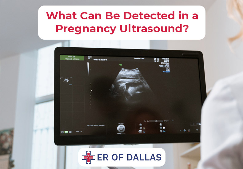
To assist medical professionals in screening for congenital disorders, ultrasound is a crucial tool (conditions your kid is born with). One kind of test that can be used to ascertain whether your baby has a particular health condition is called a screening. When doing certain diagnostic procedures during pregnancy, such as amniocentesis or CVS (chorionic villus sampling), your practitioner may additionally utilize ultrasound to guide the needle.
To determine whether your baby is receiving adequate oxygen, a biophysical profile (BPP) test that combines ultrasound and a nonstress test includes an ultrasound as well.
What to Expect From Your First Pregnancy Ultrasound
An important aspect of pregnancy is anticipation. You’re curious about the appearance and, more importantly, the health of your unborn child. Ultrasound provides an early look inside the womb and an opportunity to find out more about the health and expected date of your unborn child.
Typically, a first-trimester ultrasound is performed seven to eight weeks after the start of your most recent menstrual cycle. Your doctor can also use this test to screen for genetic problems, as well as to find any issues with your uterus or cervix. If you’re anxious to learn the baby’s sex, you’ll have to wait a bit longer. The gender reveal, as well as more info about your baby’s anatomy, will come at your next ultrasound, which happens between weeks 18 and 22 of your pregnancy.
An image of your baby’s cross-section in two dimensions is produced by a standard ultrasound. Certain facilities promote ultrasounds that are 3D or even 4D, which create an image of your baby that is closer to a photograph. While not required, these advanced scans might be ideal if you think your child has a condition like a cleft palate, which is more difficult to detect with 2D imaging.
One of two methods can be used for a prenatal ultrasound: transvaginally (into your vagina) or transabdominal (over your belly). If your pregnancy is very early on, you might consider getting a transvaginal ultrasound since it creates a more realistic picture of your still-tiny baby.
How to Prepare for An Ultrasound
You might need to have a full bladder for the ultrasound tech to get a good picture of the fetus and your reproductive organs early in the pregnancy. One hour before your planned ultrasound, you should consume two to three eight-ounce glasses of water. It’s best not to urinate right before your ultrasound so that your bladder is full when you get there.
What Happens During An Ultrasound
You lie down on a bed or examining table for an ultrasound. A specific gel is applied to your abdominal and pelvic area by an ultrasonography professional. Since the gel is water-based, neither your skin nor your clothes should be stained by it. The gel facilitates appropriate sound wave propagation. The technician will then apply a transducer, which is a tiny wand, to your abdomen. To get black-and-white images on the ultrasound screen, they move the transducer. The image on the screen may also be measured by the technician. While taking pictures, they might instruct you to move or hold your breath. The technician then verifies that all required photos were taken and are clear. Subsequently, the technician removes the gel and empty your bladder.
What Should I Expect After a Pregnancy Ultrasound?
The gel is wiped from your tummy by your sonographer if you underwent an abdominal ultrasound. They may print off some ultrasound images for you to carry home.
Your sonographer will typically not discuss the test results with you. If your obstetrician is doing the ultrasound, they might talk to you about what they find.
If your ultrasound is done by a sonographer, an obstetrician will review the pictures and discuss their findings with you when you next see them. For same-day results, most practices arrange for your visit to coincide with your ultrasound.
Types of Pregnancy Ultrasound
When a more detailed image is needed, more sophisticated ultrasonography methods might be employed. If the doctor found issues during your standard ultrasound, these can provide the details they need to diagnose you.
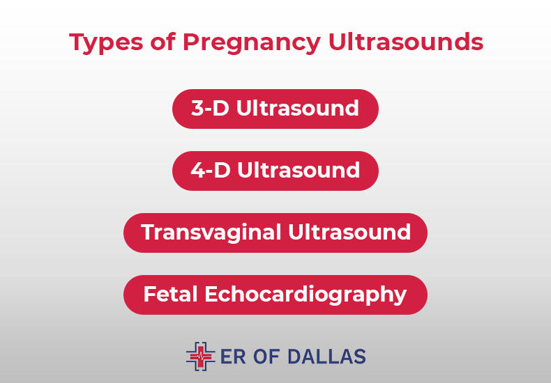
3-D Ultrasound
A 3-D ultrasound, as opposed to a conventional 2-D ultrasound, enables your doctor to view the fetus’s width, height, and depth as well as your organs. In particular, an ultrasound might be useful for diagnosing any possible pregnancy-related issues. The process for a 3-D ultrasound is the same as for a regular ultrasound; however, to produce the 3-D image, a specialized probe and software are needed. Additionally, the technician must receive specialized training, which could limit its availability.
4-D Ultrasound
Another name for a 4-D ultrasound is a dynamic 3-D ultrasound. A 4-D ultrasound, in contrast to other types, produces a moving movie of the fetus. It improves the representation of the baby’s features and motions. Additionally, it produces greater shadows and highlights. Similar to other ultrasound procedures, this one is carried out with specialized equipment.
Transvaginal Ultrasound
For a sharper view, a transvaginal ultrasound may be performed. Early in a pregnancy, when it may be harder to get a clear image, is when this ultrasound is most likely to be used. A tiny ultrasonic probe is placed into the vagina for this test. While the pictures are being taken, the probe is pressed up against the rear of your vagina.
Fetal Echocardiography
In the event that your doctor believes your child may have congenital heart abnormalities, a fetal echocardiogram is ordered. This test might take longer to finish, but it might be performed similarly to a conventional prenatal ultrasound. It takes a detailed picture of the fetus’s heart, displaying its dimensions, form, and composition. Your baby’s heart’s functioning is also seen by your doctor during this ultrasound, which can aid in the diagnosis of heart issues.
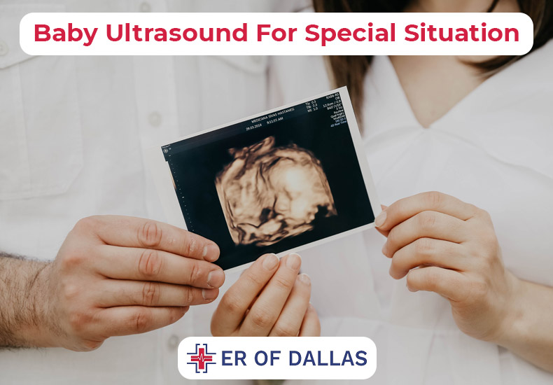
Baby Ultrasound For Special Situation
A prenatal ultrasound may be advised by your healthcare professional in circumstances other than those listed above. For instance, if you have a surgery that involves ultrasound guidance or if you have special health concerns that call for particular monitoring, ultrasounds may be recommended.
Doppler Ultrasound Pregnancy
A unique imaging test called Doppler ultrasound can be used to see blood flowing through vessels. A Doppler ultrasound during pregnancy can assist assess the health of your unborn child’s blood circulation. Doppler ultrasound in high-risk pregnancies may lower the incidence of perinatal death and obstetric interventions, according to a Cochrane analysis.
Fetal Doppler ultrasonography may be suggested by your healthcare practitioner in the following situations:
- You suffer from diabetes.
- Your blood pressure is elevated.
- You suffer from renal or cardiac issues.
- There is improper placental development.
- suspected issues with fetal growth
Additionally, the Doppler technique is used by handheld fetal heart rate monitors. These devices are frequently used by medical professionals to keep an eye on your baby’s heartbeat during birth and prenatal examinations. The FDA warns against using devices at home owing to a lack of control and needless ultrasound exposure, even though they are available over the counter.
Guidance Ultrasound
Your physician might also prescribe additional pregnancy Ultrasound tests that need direction from ultrasounds. These could include amniocentesis or chorionic villus sampling (CVS), which screen the fetus for congenital abnormalities. Ultrasound technology is also used in fetal echocardiograms, which display the baby’s heart rate and identify defects.
How Many Ultrasounds Do You Have During Your Pregnancy?
Most expectant mothers undergo one or two ultrasounds throughout their pregnancy. The quantity and time, however, may according to your healthcare practitioner during pregnancy and any underlying medical issues. Your doctor may advise more frequent ultrasounds if your pregnancy is high-risk or if they think you or your unborn child may have a medical condition.
How Early Does Ultrasound Detect Pregnancy
An embryo can be found on an ultrasound as early as week six of pregnancy, according to pregnancy care specialists. A fetus begins to develop from an embryo around the 8 weeks of pregnancy forward.
It can be too early to detect a fetal heart rate if your most recent menstrual cycle isn’t correct.
How To Read Pregnancy Ultrasound Report?
You can better understand your doctor and your pregnancy ultrasound report by following these steps:
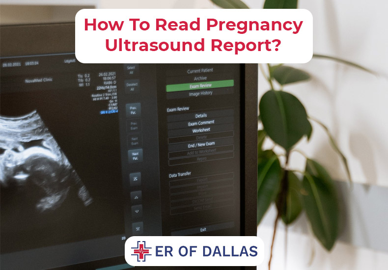
Take a Look At Your Womb
There will be a thin white or light gray border around the corners of the image when you look at your womb during the ultrasonography. Amniotic fluid is the huge black region visible inside these lines.
Take a Look At Your Baby
In the dark region mentioned above, the amniotic fluid will make your baby appear gray or white.
- Your fetus will resemble a baked bean in size and shape when it is seen via ultrasound during the first eight weeks of pregnancy.
- You will see the baby’s head if you have a scan at 12 weeks.
- There will be a noticeable difference in the scan at 20 weeks. You will be able to detect the feet, heart, spine, and eyes of the infant.
Are The Number On The Ultrasound Picture Relevant To You?
Your name, hospital reference codes, machine calibration settings, and other pertinent data recorded by technicians are represented by the digits at the top of your scan report. The tissues above your uterus will be seen in the topmost ultrasound image that the technician takes when the exam begins. The ultrasound imaging will also show you your uterus’s lining, inner side, and rear.
How Much Does Pregnancy Ultrasound Cost?
The location of the ultrasound, the body part being examined, and the type of ultrasound machine utilized can all affect the cost of the scan. The Pregnancy ultrasound can cost anywhere between $200 and $800 on average. Where you reside might also have a big impact on the pricing.
Where Can I Get An Ultrasound Near Me?
For Pregnancy ultrasound services in Dallas, I recommend checking the ER of Dallas. Contact them directly through their official website or by calling the hospital’s main line to inquire about the availability of ultrasound services and to schedule an appointment. Ensure to discuss your specific imaging needs with the ER staff for personalized guidance and information.
Conclusion
Pregnancy-related ultrasounds can be both thrilling and worrisome. Ultrasound helps your prenatal care provider better understand the growth and development of your unborn child. Ultrasounds come in a variety of forms, and the precise time will depend on your provider. The majority of pregnant women undergo two ultrasounds: one during the first trimester and one during the second. However, your provider will request further ultrasounds as a precaution if there is a possible issue or a medical need for them. Discuss with your healthcare at the ER of Dallas for Pregnancy ultrasound during your pregnancy and what to anticipate.