

ECG, is a well-known basic diagnostic method widely used in medical care for the ECG, or electrocardiography, to evaluate the heart’s electrical and muscular functions. An ECG being equipped with technology to outstandingly perceive the heartbeat is the feature that differentiates it from other monitoring techniques and explains its ability to show anomalies from minor to lethal diseases accurately.
This paper is intended to explain the fundamentals behind an ECG by discussing its major sides in detail. First of all, we will describe the ECG in general by telling what ECG is. Then, we won’t skip over how the ECG works. And finally, we will show the parts of the ECG. Making use of this procurement of knowledge, a detailed account of how to read an ECG will be provided, differentiating between normal and abnormal readings. Moreover, this working session will be an opportunity to make clear terms like ‘sinus rhythm’ and ‘borderline ECG; their meaning and implications to the clinical practice.
What is an ECG?
ECG or electrocardiogram is a noninvasive test that draws a chart of heart rhythm using electrodes placed on the skin during a period. Those electrodes then sense the smallest electrical changes that occur on the surface which are due to the heart muscle’s electrophysiologic pattern of depolarization and repolarization of each heartbeat. In brief, the ECG figuratively draws a picture of the electrical current activity of the heart which can be very useful for doctors to predict various cardiac diseases.

How Does an ECG Performed?
ECG or Electrocardiography machine is the entire system composed of many functional parts such as leads, electrodes, and data recording systems. The standard ECG has 12 leads, each one made differently for observation of the heartbeats. These leads were selected from six skeletons followers and six precordial (chest) ones. The electrodes, in a usual case, there are ten of them, are located on particular sites of the patient’s arms, legs, and chest. The electrical activity of those electrodes would be later transformed into rhythmic waveforms. Thus generated signals appear as fine lines on the ECG paper and are later interpreted by any healthcare professional to identify changes if there are any.
Components of an ECG
- P wave: Demonstrates impulse arising from atriums.
- QRS complex: Symptoms of tachycardia, bradycardia, prolonged cardiac arrest, and slow development of atrial fibrillation are seen.
- T wave: Indicates repolarization of the ventricular. In addition, you will discuss how to prevent and manage the immediate effects of cardiac arrest.
- U wave: This phenomenon occurs when a wave reaches a transmitting antenna and is seen and then represents further repolarization.
How to Read ECG?
Accuracy in the examination of an ECG, or ‘electrocardiogram’, is key to the diagnosis of heart defects and the management of cardiac health. This part contains instructions on how to understand basic ECG components by directing a user through the readings.

Knowing ECG Paper and Measures
The stationary ECG strip now reads the electrical impulse changes that occur in the human heart on graph paper which is divided into small squares, using a standardized measurement. Every little square has its unique coordinates representing 0.04 seconds at its horizontal axis and 0.1 millivolts taking the vertical axis. The bigger squares are composed of 5 squares and 5 small squares and stand for the time of 0.2 and 0.5 millivolts respectively. Identifying these references that help in measuring the pulse and heart’s electrical signals is fundamental to analyzing the rhythm.
Identifying ECG Components
- Rate: Identify the number of QRS complexes within a set time or work it out by the count of large squares between the start of a wave and the next similar one and then dividing this by 300.
- Rhythm: Identify whether the character of the heartbeat is regular or irregular. This could be achieved by measuring the time intervals between successive QRS complexes on the ECG.
- Axis: One can find out the orientation of the heart’s electrical axis by examining the lead of QRS complexes which are going toward a specific direction. It indicates if the electrical activity is normal or shifted because the cause of the abnormality is hypertrophy or conduction of the heart muscle movement is slowed down or blocked.
- Intervals: For this purpose, time the lag between the waves.
- PR interval: Detects the using atrial contraction and conduction.
- QRS duration: It marks the conduction pathway time.
- QT interval: In the duration of ventricular systole and diastole.
Steps to Read a Basic ECG
- Check Calibration and Patient Info: It is very important to make sure always that the ECG is accurate to a standard size (10mm/mV ), check the patient’s information after which take the ECG.
- Determine Heart Rate: Perform the R-R interval method of heart rate computation.
- Assess Rhythm: Establish a link between the P waves and QRS complexes to ensure you provide consistent information.
- Evaluate Waveforms: Interpret the characteristics of each P wave, QRS complex, and T wave concerning the dimensions, duration, and morphology.
- Identify Possible Abnormalities: Search for any deviations away from the typical wave pattern which could be extra beats, skipped beats, or waveform abnormalities.
What Does a Normal ECG Look Like?
Consequently, the findings of a standard ECG (ECG) display equal interval peaks with every heartbeat, which is the sign of a heart in a normal condition. It features a sequence that is rhythmically consistent where the P wave comes before the QRS wave and then the end line is the T wave beating. On the ECG tracings, the P wave represents atrial depolarization, the QRS complex implies ventricular depolarization, and the T wave signifies ventricular repolarization. This part must be symmetric and with a consistent spatial gap between them like they reflect the heart’s predictable regular beating.

How To Check if ECG Report is Normal?
To determine if an ECG report is normal, several key aspects must be evaluated:
- Heart Rate: The LHR ( Hart Rate) In normal condition it is about 60 to 120 beats per minute.
- Rhythm: It is a cyclic movement of P wave and QRS wave. Each P wave should be followed by a respective QRS wave.
- Intervals and Segments: Look out that the PR interval range is between 0.12 and 0.20 seconds, the QRS duration is below 0.12 seconds, and the QT duration is about a heart rate.
- Waveforms: Also ensure that all leads show normal P, QRS waves, and T waves without significant deviations.
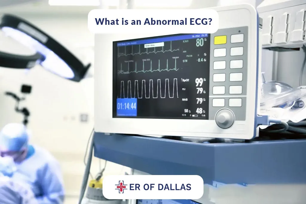
What is an Abnormal ECG?
An abnormal ECG differs from a regular rhythmic and predictable pattern. It may manifest as a nightmarish heartbeat such as too fast (tachycardia), too slow (bradycardia), or irregular patterns that often indicate arrhythmias. The magnitude and shape of the waveforms, involving P waves widening, QRS complexes elongating, and T waves becoming distorted is taken as the symptom of heart enlargement, ventricle hypertrophy, and myocardial ischemia. It is crucial to discover these dissonances as they may be attributed to the presence of underlying heart disorders that need to be subjected to proper check-ups and treatment.
What is Sinus Rhythm in ECG
The natural pacemaker, that is, the SA node, is the site where the sinus rhythm arises from, the latter being the controlled, synchronous, and regular heart rhythms. Sinus rhythm awareness is of paramount importance in that it provides benchmarks against which deviation caused by heart abnormalities can easily be noticed.
The SA node is situated in the right atrium of the heart where these electrical impulses are responsible for initiating every heartbeat with a sinus rhythm. These excitatory trainers are what trigger the atria contractions that create the P wave.
Characteristics of Sinus Rhythm
- P Wave: In total, QRS complexes are preceded by P waves. The shape of the P wave in leads I and II, as well as lead aVI, is upright and reversed for the aVR lead.
- Regular Rhythm: The intervals between these heartbeats (R-R intervals) are also what is most constant about ECGs.
- Rate: Normally, the sinus rhythm impulse is between 60 and 100 beats per minute at rest.
- P-R Interval: “It involves a constant period from the start of P wave till the start of QRS complex with the duration ranges from 0.12 to 0.20 seconds”.
ECG Remarks on How to Identify Normal Sinus Rhythm.
To confirm that the heart is in sinus rhythm, the pattern of the ECG will be regular and consistent with similar-looking P waves at the centers of the ECG. Further, every P wave must be followed by a QRS complex. Heart rate by choosing the proper Pulse Wave which should be inside a normal range and with PQ interval once it has been established.
Importance of Sinus Rhythm
A healthy sinus rhythm means that the heart’s electric system is working itself out as normal. It is the one a rhythm and underlines that atria and ventricles are dealing with the normal regularity without disturbance, and obstruction. Anomalies from sinus rhythm, like sinus tachycardia and sinus bradycardia, may speak of preexisting health problems that determine the obtention of heart rate in line with the normal condition.
What is The Difference Between EKG and ECG?
These terms, ECG and EKG, do not carry a difference, as both are designed to measure the heart’s electrical activity in a certain amount of time. This part though can be confusing since the usage of two different abbreviations requires clearing up. Affirming that both phrases have the same meaning and sometimes it makes no difference by which one you call the test, should help deal with this problem.

Origin of the Terms
- ECG: The speaker, by using this acronym, regardless of its English interpretation, is referring to an electrocardiogram.
The root of the word electrocardiogram is from the Greek language, meaning: elektron (amber), kardia (the heart), and writing (gram).
- EKG: This abbreviation is derived from the German word “Elektrokardiogramm,” which is written with ‘kardo’ spelled with a K. ‘The detector in the graph was labeled ‘K,’ which is a common use of ‘K’ in the German language and it became quite popular in the early stages when people started cardiac testing.
Usage of ECG vs.EKG
In the US, most people prefer the term EKG instead of ECG because it may confuse one with EEG (electroencephalogram) which is a test that can measure brain waves. The separation is useful between healthcare providers and the patients avoid a clash in perceptions when diagnostic tests are mentioned and touched on instantly.
Where we live, once English has become the language of health practice, accidentally, “ECG” takes the lead.
Lack of Difference in the Test
Be it ECG or EKG; a test no matter what is called is still the same. The test is performed by putting the electrodes on the patient’s chest and limb to detect and display the heart rhythm and its functions by graphs.
Since ECG and EKG are two names of the same test, standardization of the terminology in the international medical education field enhances global medical communication. The health care providers trained in a specific area will have to learn about the same names of drugs for the same purposes irrespective of the geographical regions. This will help them to communicate effectively as they traverse borders.
What is Borderline ECG?
The literal word for “Borderline ECG” would be “Near failure ECG”, and it means the cardiogram reading is near the limits. It may also indicate the upcoming failure of the cardiac system.

An ECG, which forms a boundary of normal ranges, it may show results that are on the edge of normal representations or that are unremarkable as they are insignificant from the point of view of a pathological condition. The alterations low in the hierarchy might be simple changes in heart rate, small deviations in PR, QRS, or QT intervals, or the forms of the waves being mildly abnormal in shape.
What Does Borderline ECG Mean
“Borderline ECG” as a phrase that “can seem confusing for both patients and health care providers” can, thus, trigger the need to elaborate on it further. It means there is a record of an ECG that shows findings that are neither conclusively normal nor abnormal. It is used when a patient complains of symptoms but cardiac disarrays included in the ECG report cannot be identified. Knowing what abnormal borderline ECG is indicative of timely actions and cogent management is an essential point.
Three Possible Causes of a Borderline ECG.
- Variations in Normal Anatomy: Small anatomical changes could result in unique ECG waveforms that are normal but seem aberrant.
- Electrode Placement Errors: The wrong ECG lead positioning could alter the original ECG readings and not provide a clear indication of an underlying heart condition.
- Physiological Factors: Factors involving the ability to keep a normal blood pressure level, normal levels of electrolytes in the blood, or increased heart rate (coming from anxiety, caffeine, etc.,) may change ECG readings temporarily.
Clinical Implication of Borderline ECG
Interpretation of ECG that crosses into the gray zone demands close examination and also additional tests to verify if the concerning results are clinically relevant. The approach may include:
- Repeat ECGs: To know if the flight attendant changes are permanent or if the staff returns to standard practices.
- More Comprehensive Testing: For instance, echocardiography or stress testing to have your heart precisely evaluated concerning its function and structure.
- Monitoring: For many cases, a regular follow-up would be advised if structures dictate a heart problem development.
How is An ECG Performed on a Woman?
Women’s ECG service is all about doing it with high exactness and care for her needs for sure. The opening phase of the procedure is focused on making precise saturation of the electrodes at the right locations are taken into account. They must be located lower beneath the breast to a non-coping position with interfering electrical signals generated from the mammary glands which could result in scene unrealistic imaging. The skin that will be penetrated should be freshly cleaned and hairs must always be trimmed to guarantee complete contact. The testing should be marked by contact throughout the process.

The patient should be positioned comfortably, left partially covered, with a gown or sheet, and instructed to remain calm. Breathing normally shouldn’t present any issue for them. For this reason, an EEG electrode sits as “still and undisturbing” as efficiently as possible to stop any muscle tremors or movement that could affect signal recording. Ongoing monitoring by a medical professional serves as the “golden rule” for it helps to compare the clear ECG trace with the other data and correctly read them with the help of a professional who is well-versed in their features and peculiarities.
Understanding and taking into consideration the reviewing the organismal differences such as hormonal fluctuations which can affect cardiac operability is necessary for women. Endogenic hormone fluctuations that occur around menstrual cycle phases or treatment options such as hormone replacement therapy can alter both specific features of the ECG. bearing in mind these aspects during both the evaluation of ECG and its analysis ensures a more accurate depiction and better control of the patient’s cardiac health.
Conclusion:
Electrocardiograms (ECG) serve an instrumental role in the credible diagnosis of heart diseases. This guide has enlightened you on the signing and deciphering of ECG readings, therefore helping you better understand and interpret the same. Dallas residents and visitors who are seeking more information or specialized heart-grand evaluation should feel free to visit the ER of Dallas for ECG/EKG. Their group of professionals is going to eliminate all severe treatments and give you comprehensive cardiac care and assessments.
FAQs
1. What is an ECG and for which purposes it is indicative?
ECG is an electrocardiogram that is a medical test that records the rhythm of the electrical activity of the heart continuously by using the electrodes on the skin surface. It can be used for this purpose in monitoring the health of the heart and can expose problems such as arrhythmia, heart attacks (myocardial infarction), and other cardiac diseases.
2. Will It be Appropriate Preparation for an ECG?
As for the ECG, the preparation usually you are expected to do yourself would be minimal. You should don a lightweight, soft garment that can be tidied up to place electrodes in place. Do not use oily or moisturizing skin products on the day of the test for the presence of the electrodes to your skin. Besides, it is necessary to be calm and refrain from doing exercise or taking coffee an hour then the test.
3. Is an ECG procedure comparable to a term or painful?
ECGs are not painful. Besides getting some minor discomfort from removing the adhesive you will probably get from taking a band-off, the test itself is bloodless and does not squeeze electricity. It just records the activity taking place within your body and does not produce any.
4. How long is an ECG and what should I do to prepare for it?
As the procedure is said to take between 5 to 10 minutes, it is quite convenient. What one may see (on an EKG) as a line with many wavy elements for a few seconds represents the electrical activity of the heart in its true time frame. While the patient being monitored is up for the rest of the time, the clinic’s job is to prepare, as, arrange the electrodes.
5. Is it safe for me to go back to normal activities immediately after the ECG test?
Yes, if the pathophysiological effect of the ECG is normal, you can resume your normal activities immediately unless advised otherwise by your doctor. The test will create no complications that could emerge after the research so you won’t have to fear anything.